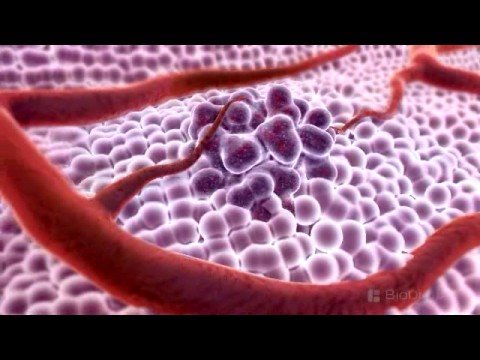
cancer (medical term: malignant neoplasm) is a diseas in which a group of cells display uncontrolled growth division beyond the normal limits,intrusion on and destruction of adjacent tissues, and sometimes spread to other locations in the body via lymph or blood. These three malignant properties of cancers differentiate them from benign tumors, which are self-limited, and do not metastasize. Most cancers form a tumor but some, like leukemia, do not. The branch of medicine concerned with the study, diagnosis, treatment, and prevention of cancer is oncology.
cancer affects people at all ages with the risk for most types increasing with age. Cancer caused about 13% of all human deaths in 2007(7.6 million).
Cancers are caused by abnormalities in the genetic material of the transformed cells. These abnormalities may be due to the effects of carcinogens, such as tobacco smoke, radiation, chemicals, or infectious agents. Other cancer-promoting genetic abnormalities may randomly occur through errors in DNA replication, or are inherited, and thus present in all cells from birth. The heritability of cancers is usually affected by complex interactions between carcinogens and the host's genome.
Genetic abnormalities found in cancer typically affect two general classes of genes. Cancer-promoting oncogens are typically activated in cancer cells, giving those cells new properties, such as hyperactive growth and division, protection against programmed cell death, loss of respect for normal tissue boundaries, and the ability to become established in diverse tissue environments.Tumor suppressor genes are then inactivated in cancer cells, resulting in the loss of normal functions in those cells, such as accurate DNA replication, control over the complete cell cycle, orientation and adhesion within tissues, and interaction with protective cells of the immune system.
Definitive diagnosis requires the histologic examination of a biopsy specimen, although the initial indication of malignancy can be symptomatic or radiographic imaging abnormalities. Most cancers can be treated and some cured, depending on the specific type, location, and stage. Once diagnosed, cancer is usually treated with a combination of surgery, chemotherapy and radiotherapy. As research develops, treatments are becoming more specific for different varieties of cancer. There has been significant progress in the development of targeted therapy drugs that act specifically on detectable molecular abnormalities in certain tumors, and which minimize damage to normal cells. The prognosis of cancer patients is most influenced by the type of cancer, as well as the stage, or extent of the disease. In addition, histologic grading and the presence of specific molecular markers can also be useful in establishing prognosis, as well as in determining individual treatments.
For a start, it has been proven that germs and viruses do not cause cancer. Present cancer research is working on the supposition that cancer is caused by toxins of various kinds which one way or another find their way into the body and in some locations cause damage to the nucleus of a cell, turning it into a cancer cell. The presumption is that a gene within the DNA structure of the cell becomes altered so that the cell begins to multiply. Such a change to the cell's DNA is called a mutation. Also suspected of being able to cause cancer in this way are various forms of radiation, and any substance or influence that either causes cancer or tends to cause cancer is called a carcinogen. This hypothesis is a simple one, and if it were true it should be easy to prove, but although it is well known that certain substances can be carcinogenic, they are not always so, and the mechanism by which they are supposed to work has never been demonstrated. The hypothesis therefore remains only guesswork, and in the minds of most doctors cancer remains a mystery.
However, there is a theory on the causation of cancer which has been proven--one which accords to all known facts and biological laws and has been demonstrated in the laboratory. This theory, called the De-differentiation Theory of Cancer, developed from knowledge accumulated about how aerobic cells generate energy by the respiration of oxygen, and is best explained in the books of four of the 20th Century's greatest medical scientists: Dr Otto Warburg, Dr Max Gerson of Germany, Dr William F. Koch of the USA, and Dr Cornelius Moerman of Holland.
Dr Dean Burk(1904-1988) was a foundation member of the US National Cancer Institute and former head of its Cytochemistry Department. For his work in cancer research he received honors from France, Britain, Germany and Russia. Formerly Associate Professor of Biochemistry, Cornell University, he worked in cancer research at the Kaiser Wilhelm Institute in Germany and at the USSR Academy of Science, Moscow. Dr Burk was the recipient of the Domagk Prize for cancer research, a Knight Commander of the Medical Order of Bethlehem, and a Knight of the Mark Twain Society. He was co-author of the booksCancer, Approaches to Tumor Chemotherapyandcell chemistry and author of over 250 published scientific papers. Dr Warburg said Burk's outstanding and decisive discoveries in cancer research were:
1.the metabolism of the regenerating liver (1941);
2.that the malignancy of cancer was proportional to the fermentation rate of the cells (1956); and
3.that in vivo growing hepatomas produced in vivo by carcinogens were similarly more malignant the higher the fermentation rate (1964).
Dr Max Gerson(1881-1959) was Jewish but didn't enjoy Warburg's standing. He was forced to flee Germany in 1933, spending the last twenty odd years of his life working in the USA. Best known for his successful dietary treatment of migraine, lupus, tuberculosis, diabetes and cancer, Gerson was the author of more than fifty published research papers and four books. Whereas Warburg was supported and honored for his full-time research, Gerson's great work was ignored by the medical establishment. He worked alone devoting his efforts mainly to the treatment of his patients, most of whom had been given up as hopeless cases by other doctors. Under Gerson's care, Dr Albert Schweitzer, double Nobel Laureate, completely eliminated his diabetes and Schweitzer's wife her tuberculosis using Gerson's dietary methods. After Gerson's death Dr Schweitzer said of him: "I see in him one of the most eminent geniuses in the history of medicine." Notwithstanding recognition by such medical greats as Dr Schweitzer and Dr Ferdinand Sauerbruch, Gerson's work received no recognition by the medical establishment, which considered him an unorthodox threat to the medical system.
It is important to know what some of the general (non-specific) signs and symptoms of cancer are, but remember that having any of these does not mean that you have cancer -- there are many other conditions that can cause these signs and symptoms, too.
-Unexplained Weight Loss
-Fever
-Fatigue
-Pain
-Skin Changes
Specific Cancer Signs and Symptoms
Along with the above general symptoms, you should watch for the following common symptoms, which could be an indication of cancer. Again, there may be other causes for each of these, but it is important to bring them to your doctor’s attention as soon as possible so that they can be investigated.
-Change in Bowel Habits or Bladder Function
-Sores That Do Not Heal
-Unusual Bleeding or Discharge
-Thickening or Lump in Breast or Other Parts of the Body
Your bladder is a hollow, muscular, balloon-like organ that collects and stores urine. Urine is produced by your kidneys and consists of water and waste products. Tubes carry urine from your kidneys to your bladder (through your ureters) and then to the outside (through your urethra).
Your bladder is lined with a membrane (urothelium) that stops urine being absorbed back into your body. The cells of this membrane are called transitional cells or urothelial cells.
In the UK, bladder cancer is the fourth most common cancer in men and the eleventh most common cancer in women. Around 10,000 people in the UK are diagnosed with bladder cancer each year. It's rare in people under 40, but the rate rises with age.
Types of bladder cancer
There are several different types of bladder cancer. They are named after the type of cells they first occur in:
·transitional cell carcinoma (TCC)
·
squamous cell carcinoma (SCC)
·adenocarcinoma
TCC is the most common type of bladder cancer in the UK.
Some bladder cancers form small mushroom-like growths on the lining of the bladder. These are called papillary cancers.
Bladder cancer is also classified according to how far it has spread.
·Non-muscle invasive cancer - the cancer is only in the bladder lining.
·Muscle-invasive cancer - the cancer has spread to the muscle wall of the bladder.
·Advanced cancer - the cancer has spread through the bladder wall into nearby organs such as the prostate gland, vagina, bowel, or lymph nodes. Further spread to other organs such as bones and liver is possible.
Around eight out of 10 of bladder cancers are non-muscle invasive.
Symptoms of bladder cancer
Blood in your urine (haematuria) is the most common symptom of bladder cancer. This may come and go and is often painless.
The following symptoms aren't always due to bladder cancer but if you have any of these or notice blood in your urine, you should visit your GP:
· a burning feeling when passing urine
·a need to pass urine frequently
·feeling the need to urinate but not being able to
·pain in your pelvis
·recurrent urinary tract infections
·blood clots in your urine; these may cause pain
Causes of bladder cancer
The causes of bladder cancer aren't fully understood at present. However, you're more likely to develop bladder cancer if you:
·smoke - you're three to four times more likely to develop bladder cancer and passive smoking may also increase your risk
·have been exposed to certain industrial chemicals (eg in the rubber, paint, dye, printing and textile industries, gas and tar manufacturing, iron and aluminium processing)
·have had a long-term infection with the tropical disease bilharzia
·have had a long-term or repeated bladder infection
Diagnosis of bladder cancer
Your GP will ask you about your symptoms and examine you, and may also ask you about your medical history. He or she may test your urine with a 'dipstick' to look for blood and signs of an infection. Your GP may refer you to a urologist (a surgeon who specialises in identifying and treating conditions of the urinary system) for further tests, such as those listed below.
Flexible cystoscopy
This is done under local anaesthetic. It allows your surgeon to look inside your bladder using a flexible, tube-like telescope with a camera at the end, called a cystoscope. This is inserted into your urethra. If your surgeon sees anything unusual in your bladder, he or she may ask you to return for a repeat flexible or rigid cystoscopy.
Rigid cystoscopy
This is done under general anaesthetic. It allows your surgeon to pass surgical instruments into your bladder and take tissue samples (biopsies) and, if necessary, remove growths. Tissue samples are examined in a laboratory to see whether cancer cells are present and, if so, what kind of cells they are.
Intravenous urogram (IVU)
This test examines your urinary system by injecting a dye into a vein in your arm - this is gradually removed from your blood by your kidneys. By watching the movement of the dye on an X-ray screen, your surgeon can see anything unusual in your urinary system.
Scans
Ultrasound, CT (computed tomography), and MRI (magnetic resonance imaging) can help your surgeon see how far the cancer has spread (if at all).
Treatment of bladder cancer
Treatment of bladder cancer depends upon the type of cancer and how far it has spread.
Treatment of non-muscle invasive bladder cancer
Transurethral resection of a bladder tumour (TURBT)
Your surgeon will use a cystoscope and "snip off" the tumour at the stem and seal the area to prevent bleeding. The procedure takes 20 minutes to one hour and is done under general anaesthetic. Non-muscle invasive tumours often come back so you will need to have regular check-ups. See Related topics for more information.
Intravesical chemotherapy and immunotherapy
Chemotherapy uses medicines to destroy cancer cells. In intravesical chemotherapy, medicines are placed directly into your bladder using a fine tube (catheter) inserted into your urethra. This is done immediately after your surgeon has removed a tumour using TURBT. Your surgeon may repeat the intravesical chemotherapy at weekly intervals, usually for six weeks. See Related topics for more information.
Immunotherapy uses your body's immune system to fight cancer cells. The Bacille Calmette-Guérin (BCG) vaccine (used to prevent tuberculosis or TB) has been shown to be effective for treating some non-muscle invasive bladder cancers. It's put directly into your bladder (intravesical BCG) using a catheter. Treatment is given at weekly intervals, usually for six weeks. See Related topics for more information.
Treatment of muscle-invasive bladder cancer
Major surgery to remove the whole bladder and surrounding tissues is usually required (complete or radical cystectomy). Your surgeon will create a new way for you to store your urine and there are various types of operation to do this.
Urostomy
Your surgeon connects your ureters to a small opening (a stoma) in your abdomen (tummy) using a short piece of your small bowel. A flat, watertight bag is placed over the stoma to collect your urine.
Continent urinary diversion
Your surgeon makes a pouch inside your abdomen to collect urine using a section of your stomach or intestine. He or she will connect this to the outside of your body via a stoma which is kept closed with a valve. You will need to empty the pouch four to five times a day by inserting a catheter into the stoma.
Bladder reconstruction
Your surgeon may be able to make a new bladder using part of your bowel. Your urine drains from your ureters into the new bladder. You will need to learn how to pass urine through your urethra by using your muscles. You will have lost the nerves that tell you when your bladder is full and so will need to remember to empty it.
Radiotherapy and chemotherapy
Radiotherapy uses radiation to destroy cancer cells. A beam of radiation is targeted on the cancerous cells, which shrinks the tumour. Radiotherapy may be used instead of surgery.
Intravenous chemotherapy (into your vein) may be given if the cancer has spread into the muscle of the bladder. It may be given to shrink the tumour before surgery or radiotherapy treatment, or to reduce the chances of the tumour coming back after surgery.
Treatment of advanced bladder cancer
If the cancer has spread outside your bladder, you may have chemotherapy or radiotherapy. TURBT may be performed if you're finding it hard or painful to pass urine.
Prevention of bladder cancer
Factors that make it less likely that you will get bladder cancer include not smoking and eating a diet high in fruit and vegetables and low in fat.
Living with bladder cancer
Many hospitals have stoma nurses who can help you take care of your urostomy and give you advice. Most people who have a urostomy are able to take up their previous jobs, sports activities and hobbies again. Modern stoma appliances are hardly noticeable under your clothes and shouldn't leak.
Being diagnosed with cancer can be distressing for you and your family. An important part of cancer treatment is having support to deal with the emotional aspects as well as the physical symptoms. Specialist cancer doctors and nurses are experts in providing the support you need.
There are around 200 bones in your body. These bones make up your skeleton - the rigid internal structure that supports your body. Without bones your body would fall to the ground like jelly.
Bone is a living tissue. It's made up of a matrix of the mineral calcium and different types of cells. The cells continuously break down the old matrix and form new bone matrix. Most bones are hollow - within them is a type of soft tissue called bone-marrow that produces blood cells.
At joints such as the elbow or the knee, bones are covered with a thick rubbery tissue called cartilage. Cartilage allows smooth movement at the joints without damage to the bone.
As well as supporting the body, some bones, for example the skull and the ribcage, protect important organs from external damage.
What is bone cancer?
Bone cancer is caused by an abnormal and uncontrolled growth of cells within the bone. It can be benign or malignant.
Benign tumours aren't cancerous. They don't spread to other parts of the body and don't invade surrounding tissue.
Malignant tumours are cancerous. They spread to other parts of the body and invade surrounding tissue. This spread of cancer is called a metastasis and can form a secondary cancer in another organ.
Types of bone cancer
Bone cancer can be either primary or secondary.
·Primary bone cancer starts in the cells of the bone.
·Secondary bone cancer. This is cancer while starts in another organ of your body but has spread to the bones. This cancer behaves like the original cancer that it spread from and not like bone cancer.
This factsheet is about primary bone cancers.
There are many types of primary bone cancer. The main ones are listed below.
·Osteosarcoma. This is the most common type of bone cancer. About 150 people develop osteosarcoma every year in the UK. Children and young people between the ages of 10 and 20 are more commonly affected, but it can occur at any age. It is slightly more common in males than females.
·Ewing's sarcoma. This cancer also tends to develop between the ages of 10 and 20. Like osteosarcoma, it is slightly more common among males than females. This cancer can also occur in soft tissues in the body such as muscle.
·Chondrosarcoma. This is the second most common type of bone cancer. It is more common in adults between the ages of 40 and 60. It starts in the cartilage cells in joints.
·Spindle cell sarcoma. There are four types of bone cancer: undifferentiated sarcoma of the bone, malignant fibrous histiocytoma, fibrosarcoma and leiomyosarcoma. They all behave like osteosarcoma but are more common in adults.
Symptoms
The symptoms of bone cancer vary depending on where it develops and how severe it is. Different types of bone cancer tend to form in different areas. For example:
·osteosarcoma is most common in the lower thigh, shins and arms
·Ewing's sarcoma most commonly occurs in the pelvis, thigh and shins
·chondrosarcoma is most common in the thigh, pelvis, ribs, upper arm and shoulder bone.
·spindle cell sarcoma most commonly develops in the lower thigh, shins and arms
Bone cancer often causes pain and tenderness in the affected area. This is often worse at night. As the cancer grows it can also cause swelling in the affected area. If it is near a joint it may make movement in that area difficult.
Less common symptoms of bone cancer include:
·tiredness
·fever
·weight loss
It's important to remember that these symptoms can be caused by many problems other than bone cancer. So although not necessarily a result of bone cancer, if you have these symptoms you should visit your GP.
Causes
No one knows exactly what causes bone cancer, but research is ongoing. But there are some things that increase your chances of getting bone cancer.
·Previous treatment with radiotherapy. If you have had a lot of radiotherapy for cancer in the past, you have a slightly increased risk of getting bone cancer in that area.
·Paget's disease. This bone disease gradually deforms your bones, causing pain and fractures. Having Paget's disease for a long time increases your risk of developing bone cancer
·Having a previous benign bone tumour. If you have had a benign (non-spreading) type of bone cancer, you are more likely to develop chondrosarcoma.
·Retinoblastoma. Inheriting the gene that causes this rare type of eye cancer also makes you more likely to develop osteosarcoma.
·Having certain other rare inherited conditions, such as Li-Fraumeni syndrome, can increase your risk of developing bone cancer.
Diagnosis
Bone cancer can be diagnosed by many different tests.
X-ray
Doctors are often able to diagnose bone cancer from an X-ray image of the affected area. X-rays can sometimes be useful for finding out the type of bone cancer it is.
Bone scan
For having a bone scan a small amount of harmless radioactive dye is injected into a vein. This collects in areas of the bone that may have cancer, and is picked up by the scan.
Bone scans are better than X-ray images at showing up a bone cancer. But other diseases, such as arthritis, can also cause a positive result. So if you have a positive bone scan, you may need further tests to make sure you have bone cancer.
MRI scan
Magnetic resonance imaging (MRI) scans use magnets and radio waves to produce images of the inside of your body. Tumours growing inside bones can be seen with MRI scans.
Biopsy
Often your doctor will want to take a biopsy to check if a tumour is non-cancerous. A biopsy is a small sample of tissue. This will be sent to a laboratory for testing.
The biopsy is usually done using a long needle, under local anaesthesia. The procedure is called a core needle biopsy. Sometimes doctors do an operation called a surgical biopsy. This can be done under local or general anaesthetic.
Further tests
If you are diagnosed with bone cancer, you will have more tests to check if the cancer has spread. This is called staging. You are likely to have a chest X-ray, to see if it has spread to your lungs, or a computerised tomography (CT) scan to look for signs of cancer elsewhere in your body.
Treatment
How bone cancer is treated depends on the type of bone cancer you have, how far it has spread, your age and your general health. The treatment for these rare tumours is carried out in expert centres where cancer specialists (oncologists) and surgeons are familiar with the special treatments required. There are three main types of treatment for bone cancer.
Surgery
The type of surgery you have depends on how far the cancer has spread.
·Limb salvage surgery involves removing the area of bone where the tumour is. Because of the recent advances in surgery, this method of treating bone cancer is becoming more common. The area of bone removed is replaced with either a metal prosthesis (an artifical replacement part) or a piece of healthy bone taken from another part of your body (a bone graft).
·Despite ongoing improvements in surgical technique, sometimes a limb salvaging operation isn't possible. If the cancer has spread into surrounding tissues, amputating the limb may be the only way to get rid of the cancer. Support from the medical staff looking after you can help you come to terms with this news. Advances in prosthetics (artificial limbs) mean that you can often have a fully active life after this surgery. A specialist in artificial limbs will visit you at hospital to arrange one for you. A physiotherapist will be able to teach you how to adapt to and best use it.
Non-surgical treatments
Chemotherapy
Chemotherapy uses medicines to destroy cancer cells. However, they can also have side effects such as making you feel tired or ill, or causing nausea or hair loss. Chemotherapy is particularly good at treating Ewing's sarcoma, but it can also treat other types of bone cancer such as osteosarcoma.
There are lots of different types of chemotherapy drugs. They are usually injected into a vein but sometimes tablets are used.
Chemotherapy is often given before and after surgery to make it easier to remove the tumour and to prevent it coming back.
Radiotherapy
Radiotherapy uses radiation to kill cancer cells. A beam of radiation is targeted on the cancerous cells, which shrinks the tumour.
Radiotherapy is especially useful for Ewing's sarcoma but it's sometimes used for osteosarcoma. It can be used before surgery to make it easier to remove the tumour, or afterwards to prevent it coming back.
After your treatment
If you have had an operation you may need physiotherapy and other support to help get you back to functioning well. You will also be seen regularly by a specialist to make sure the cancer hasn't returned.
 RSS Feed